Comparison · 10/15/2026
Gefitinib, erlotinib and osimertinib: structures and binding evidence
A worked EGFR comparison that separates molecular observations from experimental binding evidence.
Read the two structures
Compare the ring regions, oxygen-containing connections and nitrogen-containing side chains of osimertinib and gefitinib in sequence. Reading regions separately makes shared and different local environments easier to describe without equating target-family membership with a shared complete scaffold.
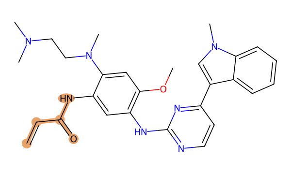
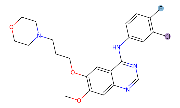
| Observation | Osimertinib | Gefitinib |
|---|---|---|
| Acrylamide connectivity | 1 | 0 |
| Fluorine atoms | 0 | 1 |
| Chlorine atoms | 0 | 1 |
Connect the structural observations with the fingerprint result
One C=C–C(=O)–N motif is detected in osimertinib and none by the same query in gefitinib. Gefitinib’s one fluorine and one chlorine are marked separately. This provides concrete connectivity and element observations within the EGFR grouping. [Osimertinib] · [Gefitinib]
Nineteen shared set bits across a union of 111 give 0.171. The class groups comparison subjects by target context, while this calculation compares radius-2 atom environments. These are different kinds of similarity.
The unsaturated connection is a structural query result. Reaction-rate and protein-binding measurements are absent, so the coloured motif cannot establish binding mode or activity against a particular mutation.
Calculated structural similarity: 0.171 · Morgan radius 2 / 2048 bits · Tanimoto
Calculation: 19 / (70 + 60 − 19) = 19 / 111 ≈ 0.171. Set-bit counts are not counts of unique atoms or functional groups.
Query definitions and reproducibility
- Acrylamide connectivity:
C=CC(=O)N - Fluorine atoms:
[F] - Chlorine atoms:
[Cl]
RDKit 2025.03.4 · Matches with identical atom sets are counted once. Colours mark atoms and bonds matching selected queries, not a proven shared scaffold or synthesis route.
- Read elemental composition from the formulas.
- Trace ring and substituent connections in each drawing.
- Compare your observations with the fingerprint score calculated under identical settings.
- Check stereochemistry and the complete InChIKey in the source records.
The score is fingerprint overlap, not a percentage of efficacy or safety. RDKit
Structure-source check: 2026-10-08 / 2026-10-08 · Editorial update and calculation check: 2026-10-16
Download source structures, queries and bit counts for this comparison (JSON)
Question · does one EGFR family imply one structure?
Gefitinib, erlotinib and osimertinib share a navigation category, not a molecular identity. Start with the two independent records below and open the third compound through the related links. The aim is to identify what a structure comparison can establish, rather than rank therapies.
Observation 1 · follow connectivity
Record the CID and formula for each molecule. Distinguish nitrogen within a ring from nitrogen in a side chain. Drawing orientation is not molecular identity. This page calculates an osimertinib–gefitinib pair; it does not claim to calculate a maximum common substructure across all three drugs.
Observation 2 · motif detection is not an experiment
Highlighted counts report matches to disclosed SMARTS queries. Finding a motif does not observe a reaction in a protein. Neither a shared motif nor a high fingerprint score establishes binding kinetics, reversibility or selectivity. Those conclusions need experimental evidence.
Experimental context · read the conditions in 4ZAU
PDB 4ZAU records an X-ray structure of wild-type EGFR with AZD9291 at 2.80 Å resolution. Check the actual sequence, mutation field, ligand and experiment before interpreting the model. A wild-type complex does not establish activity against every resistance variant.
Reproduction · match the inputs first
Download the two SMILES and CIDs, radius-2 Morgan settings, 2048-bit size and chirality setting. Divide shared set bits by union set bits to check the displayed Tanimoto. If another service differs, compare chemical species and fingerprint parameters before interpreting the number. No receptor protein or activity measurement enters this calculation.
Boundary · clinical results require other evidence
Report the structural observation with its calculation settings. Variant-specific activity, exposure, safety and patient outcomes require separate evidence. This guide is a structure-reading exercise rather than a current approval list or treatment-selection guide.
Sources and review
Editorial update: 10/15/2026 · Structure-source check: 2026-10-08. Calculations and cited experiments are separate evidence.
Mechanism classes
Related compounds
Editorial responsibility and corrections
Operated and edited by the independent StructureMoa project. Explanations use AI assistance; structural identity and similarity are checked against source records and calculation tests. Molecular data and clinical or regulatory evidence have separate review scopes.
- Source structures
- PubChem · CID / SMILES / InChIKey
- Calculations and automated checks
- RDKit · Morgan / SMARTS
- Disclosed specialist review
- No separate external specialist review is disclosed. Passing automated tests is not specialist review of clinical claims.
For a correction, include the page URL, molecule name or CID, the field in question and a supporting source. Corrections are checked against sources, and the affected editorial update date is changed when a correction is published. [email protected]