Comparison · 10/7/2026
KRAS comparisons: molecular structure and binding state
Read sotorasib and adagrasib independently, and distinguish daraxonrasib’s RAS(ON) approach.
Read the two structures
Trace the connections between rings in adagrasib and compare them with sotorasib. Similar element labels do not establish the same ring arrangement or substitution positions. The fingerprint score summarizes overlap of encoded local atom environments, not a common synthetic origin.
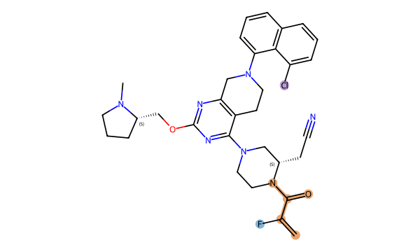
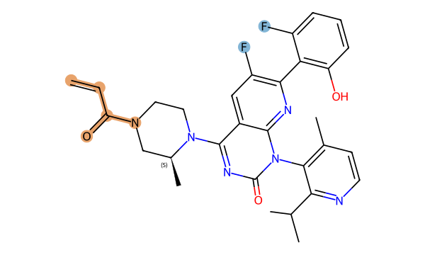
| Observation | Adagrasib | Sotorasib |
|---|---|---|
| Acrylamide connectivity | 1 | 1 |
| Fluorine atoms | 1 | 2 |
| Chlorine atoms | 1 | 0 |
Connect the structural observations with the fingerprint result
The orange C=C–C(=O)–N connectivity occurs once in each molecule. Sotorasib has two fluorine atoms attached to aromatic rings; adagrasib has one fluorine attached to the highlighted unsaturated connection and also one chlorine atom. The location and neighbouring atom environments differ, not just the element counts. [Adagrasib] · [Sotorasib]
Sotorasib has 75 set fingerprint bits and adagrasib has 83. They share 21, so the union is 75 + 83 − 21 = 137 bits. The rounded result of 21/137 is 0.153. A small common motif can coexist with limited overlap across the complete fingerprint.
The result describes radius-2 local environments in this bit representation. We have not assigned individual bits to chemical groups, so the score cannot be decomposed into a numerical contribution from fluorine or chlorine. Biological binding and efficacy require separate experimental evidence.
Calculated structural similarity: 0.153 · Morgan radius 2 / 2048 bits · Tanimoto
Calculation: 21 / (83 + 75 − 21) = 21 / 137 ≈ 0.153. Set-bit counts are not counts of unique atoms or functional groups.
Query definitions and reproducibility
- Acrylamide connectivity:
C=CC(=O)N - Fluorine atoms:
[F] - Chlorine atoms:
[Cl]
RDKit 2025.03.4 · Matches with identical atom sets are counted once. Colours mark atoms and bonds matching selected queries, not a proven shared scaffold or synthesis route.
- Read elemental composition from the formulas.
- Trace ring and substituent connections in each drawing.
- Compare your observations with the fingerprint score calculated under identical settings.
- Check stereochemistry and the complete InChIKey in the source records.
The score is fingerprint overlap, not a percentage of efficacy or safety. RDKit
Structure-source check: 2026-10-08 / 2026-10-08 · Editorial update and calculation check: 2026-10-16
Download source structures, queries and bit counts for this comparison (JSON)
Begin with the small molecules
The same target family can contain different molecular architectures. Compare the independent sotorasib and adagrasib drawings, formulas and identifiers below. A family label is a navigation aid, not proof that one molecule was synthesized from the other.
Separate molecular architecture from mechanism
Daraxonrasib’s discovery paper describes a noncovalent, multi-selective RAS(ON) tri-complex inhibitor. This is a different mechanistic comparison from the KRAS G12C OFF covalent inhibitor grouping. Follow the primary paper for the binding model; a two-dimensional structure alone cannot establish the biological binding state.
Try a three-record comparison
Open sotorasib, adagrasib and daraxonrasib using the related-compound links. Record each CID and formula. Compare the ring arrangement and labelled heteroatoms first, then read the mechanism source separately. This prevents a shared target name from becoming an unsupported shared-scaffold claim.
Where the structural comparison ends
Graph position, edge count and fingerprint similarity do not establish clinical superiority. A clinical comparison requires the relevant population, mutation, treatment context and endpoints. This guide explains the structure-reading method and does not offer treatment selection or a current approval summary.
Exercise 1 · record identity before interpreting a score
Record both CIDs, formulas and complete InChIKeys from the comparison below. Compare labelled atoms and ring connectivity before using the family name. The shared and union fingerprint bits are calculations on these input structures, not pharmacology measurements. The downloadable record preserves the inputs used for this comparison.
Exercise 2 · distinguish a molecule from a protein complex
PDB 6OIM records an experimental KRAS G12C complex covalently bound to AMG 510. Use it to examine spatial context absent from a small-molecule drawing. Read the experiment and primary citation alongside the model. One crystal structure does not describe every cellular state or every RAS inhibitor.
Write a bounded conclusion
This calculation establishes overlap between encoded atom environments. ON/OFF binding-state descriptions require mechanistic evidence. Clinical benefit in a particular population or combination remains a separate question. Keep structural observations, mechanism evidence and clinical results in separate notes rather than collapsing them into a ranking.
Sources and review
Editorial update: 10/15/2026 · Structure-source check: 2026-10-08. Calculations and cited experiments are separate evidence.
Mechanism classes
Related compounds
Editorial responsibility and corrections
Operated and edited by the independent StructureMoa project. Explanations use AI assistance; structural identity and similarity are checked against source records and calculation tests. Molecular data and clinical or regulatory evidence have separate review scopes.
- Source structures
- PubChem · CID / SMILES / InChIKey
- Calculations and automated checks
- RDKit · Morgan / SMARTS
- Disclosed specialist review
- No separate external specialist review is disclosed. Passing automated tests is not specialist review of clinical claims.
For a correction, include the page URL, molecule name or CID, the field in question and a supporting source. Corrections are checked against sources, and the affected editorial update date is changed when a correction is published. [email protected]